

“The wedge.”
The holy grail of RHC and hemodynamics
Hate it or love it, major decisions are made based on it
Whether you:
• Perform the procedure yourself
• Review someone else’s tracings
• Review someone else's report
Learn 7 tips to ensure “the wedge” accuracy x.com
The holy grail of RHC and hemodynamics
Hate it or love it, major decisions are made based on it
Whether you:
• Perform the procedure yourself
• Review someone else’s tracings
• Review someone else's report
Learn 7 tips to ensure “the wedge” accuracy x.com

BUT FIRST!
You received the following RHC report:
PA Systolic 51 mmHg
PA Diastolic 22 mmHg
PA Mean 32 mmHg
Mean Wedge 30 mmHg
Should you REPEAT one of the values :
(answer before continuing the 🧵; final answer at the end)
You received the following RHC report:
PA Systolic 51 mmHg
PA Diastolic 22 mmHg
PA Mean 32 mmHg
Mean Wedge 30 mmHg
Should you REPEAT one of the values :
(answer before continuing the 🧵; final answer at the end)
7 tips to improve wedge pressure (WP) accuracy:
1. WP SHOULD NOT be higher than PA diastolic (dPAP)
2. Evidence of distinct “a” and “v” waves
3. Presence of respiratory variability
4. Stationary catheter by fluoroscopy
5. Free flow is present within the catheter
6. Obtain a wedge oxygen saturation
7. Ensure you ONLY inflate the balloon UNTIL you obtain a wedge waveform
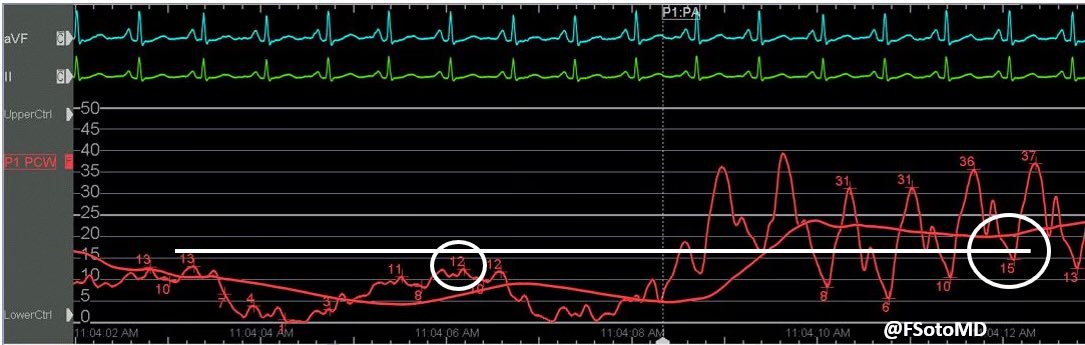
1. WP SHOULD NOT be higher than dPAP
1st image (cath lab) shows transition from WP to dPAP
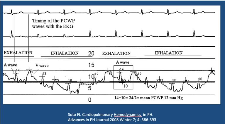
Measured at end-exhal (spontaneous breathing):
mean WP around 11 mmHg (average of the “a” waveform) and dPAP is 15 mmHg
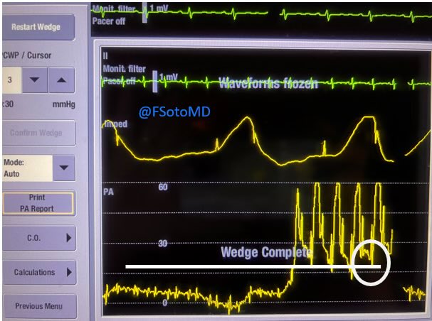
2nd image (ICU monitor) also confirms WP lower than dPAP
Image x.com
1st image (cath lab) shows transition from WP to dPAP
Measured at end-exhal (spontaneous breathing):
mean WP around 11 mmHg (average of the “a” waveform) and dPAP is 15 mmHg
2nd image (ICU monitor) also confirms WP lower than dPAP
Image x.com
2. Evidence of distinct “a” and “v” waves
Good indicator of free flow present in PA catheter:
uninterrupted column of blood between the wedged catheter and the left atrium
Will occur if the catheter tip is in West zone 3 (PA capillary pressure > alveolar pressure) x.com
Good indicator of free flow present in PA catheter:
uninterrupted column of blood between the wedged catheter and the left atrium
Will occur if the catheter tip is in West zone 3 (PA capillary pressure > alveolar pressure) x.com
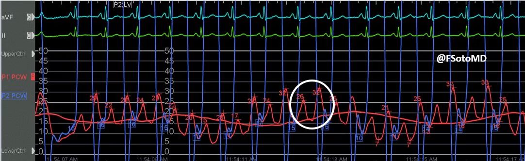
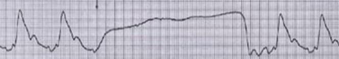
3. Presence of respiratory variability
This reassures the presence of a free, uninterrupted column and West Zone 3 location
Image 1 shows normal respiratory variability
Image 2 shows an over-wedged balloon tip (no respiratory variability) x.com
This reassures the presence of a free, uninterrupted column and West Zone 3 location
Image 1 shows normal respiratory variability
Image 2 shows an over-wedged balloon tip (no respiratory variability) x.com
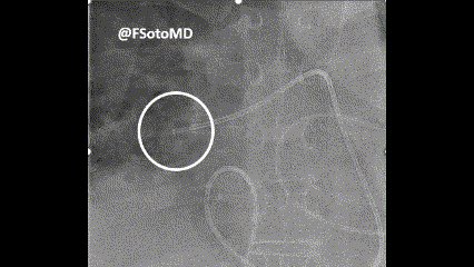
4. Stationary catheter by fluoroscopy
If RHC is performed under fluoroscopy, the tip of the catheter is “anchored” and stationary
There should be no active movement of the PAC tip into the region distal to the tip x.com
If RHC is performed under fluoroscopy, the tip of the catheter is “anchored” and stationary
There should be no active movement of the PAC tip into the region distal to the tip x.com
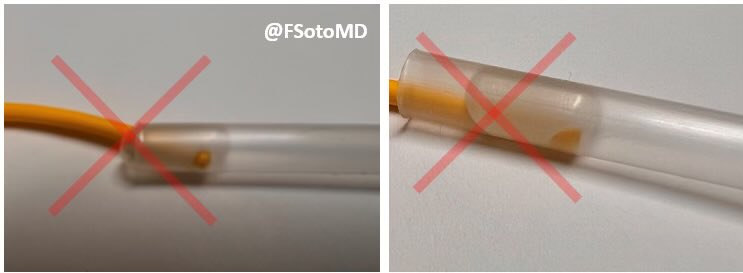
6. Free flow is present within the catheter
The 🎈 is inflated JUST ENOUGH to keep patency between PAC tip and left atrium
Image 1: over-wedged 🎈, potentially blocking the lumen
Image 2: optimally inflated 🎈 maintaining patency
Free flow allows for a successful step 6 x.com
The 🎈 is inflated JUST ENOUGH to keep patency between PAC tip and left atrium
Image 1: over-wedged 🎈, potentially blocking the lumen
Image 2: optimally inflated 🎈 maintaining patency
Free flow allows for a successful step 6 x.com
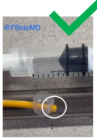
6. Obtain a wedge oxygen saturation
First, DISCARD at least 3 cc: 1 cc to remove PAC’s saline, and 2-3 cc to remove mixed venous blood from column distal to PAC tip
If 🎈 is optimally inflated, there should be free flow, drawing high O2 blood from pulm vein
Then, draw 1-2 cc with ABG syringe
If optimally placed, wedge sat should be ~ to systemic sat (👇🏻 SLOW wedge blood drawing video and picture with systemic sats)

7. ONLY inflate the 🎈 UNTIL you obtain a wedge waveform
Inflate 0.1-0.2 cc x second until wedge
See video 👇🏻
SLOW inflation technique (while looking at the monitor for change in waveform) also DECREASES risk of PA rupture x.com
Inflate 0.1-0.2 cc x second until wedge
See video 👇🏻
SLOW inflation technique (while looking at the monitor for change in waveform) also DECREASES risk of PA rupture x.com

The CORRECT answer to the test.
WHICH variable should be repeated:
• The mean WP value needs to be rechecked
• The reported dPAP was 22 mmHg, and the wedge was 30 mmHg.
• WP SHOULD NOT be higher than dPAP (can be SAME or LOWER)
WHICH variable should be repeated:
• The mean WP value needs to be rechecked
• The reported dPAP was 22 mmHg, and the wedge was 30 mmHg.
• WP SHOULD NOT be higher than dPAP (can be SAME or LOWER)
SUMMARY
7 tips to improve WP accuracy:
1. WP SHOULD NOT be > than dPAP
2. Distinct “a” and “v” waves
3. Respiratory variability
4. Stationary catheter by fluoro
5. Free flow present
6. Obtain wedge O2 sat
7. Ensure you ONLY inflate the 🎈 UNTIL wedge waveform
7 tips to improve WP accuracy:
1. WP SHOULD NOT be > than dPAP
2. Distinct “a” and “v” waves
3. Respiratory variability
4. Stationary catheter by fluoro
5. Free flow present
6. Obtain wedge O2 sat
7. Ensure you ONLY inflate the 🎈 UNTIL wedge waveform
If you appreciated this thread, please:
PLEASE
• “Like” the thread (plus individual tweets if 👍🏻)
• Repost the FIRST tweet for others to benefit from this
#FOAMed
#MedTwitter
PLEASE
• “Like” the thread (plus individual tweets if 👍🏻)
• Repost the FIRST tweet for others to benefit from this
#FOAMed
#MedTwitter
جاري تحميل الاقتراحات...