

Anatomy of the cerebellum: the arbor vitae and more. 🧠🌳
🤔🧑🏫
#Neurology #Neurotwitter #Anatomy #Teaching #EndNeurophobia
Image from Bolk's "Das Cerebellum der Säugetiere" (1906)
1/🧵
🤔🧑🏫
#Neurology #Neurotwitter #Anatomy #Teaching #EndNeurophobia
Image from Bolk's "Das Cerebellum der Säugetiere" (1906)
1/🧵

Introduction
Part of the brain in charge of integration of massive sensory and other inputs from many regions of the brain and spinal cord.
Also called "little brain" due to it's etimological origin. 🧠
2/🧵
Part of the brain in charge of integration of massive sensory and other inputs from many regions of the brain and spinal cord.
Also called "little brain" due to it's etimological origin. 🧠
2/🧵

Introduction
👷♂️ Functions?
- Motor coordination 💪👀🤹♀️
- Motor planning 🤔
- Cognitive functions (visuospatial, executive, etc)
- Speech 🗣️
3/🧵
👷♂️ Functions?
- Motor coordination 💪👀🤹♀️
- Motor planning 🤔
- Cognitive functions (visuospatial, executive, etc)
- Speech 🗣️
3/🧵

Where is it⁉️
Easy, behind the brainstem.
Connected to it throught three little feet (or peduncles) 👣
Superior 🦶
Middle 🦶
Inferior 🦶
4/🧵
Easy, behind the brainstem.
Connected to it throught three little feet (or peduncles) 👣
Superior 🦶
Middle 🦶
Inferior 🦶
4/🧵


How is it divided?
Three major ways to divide it:
Anatomical/macroscopic 🧠
Functional 🖥️
Evolutive 🦧
The last two overlap each other, therefore, let's begin with the anatomical/macroscopic.
5/🧵
Three major ways to divide it:
Anatomical/macroscopic 🧠
Functional 🖥️
Evolutive 🦧
The last two overlap each other, therefore, let's begin with the anatomical/macroscopic.
5/🧵
How is it divided?
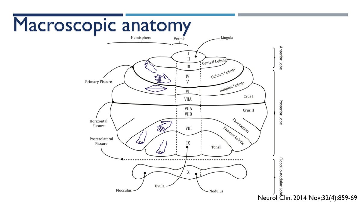
Rotro-caudal: Anterior lobe, posterior lobe and flocculonodular lobe.
Medial-lateral: vermis-hemispheres.
🚨Lobes are divided by fissures 👇
6/🧵
Rotro-caudal: Anterior lobe, posterior lobe and flocculonodular lobe.
Medial-lateral: vermis-hemispheres.
🚨Lobes are divided by fissures 👇
6/🧵

How is it divided?
Functional💻: vestibulocerebellum, spinocerebellum, cerebrocerebellum.
Evolutive🦧
Archi-cerebellum: antique 🪨
Paleo-cerebellum: not so antique🪵
Neo-cerebellum: new🛞
It will make more sense in a few tweets (promise)😕
7/🧵
Functional💻: vestibulocerebellum, spinocerebellum, cerebrocerebellum.
Evolutive🦧
Archi-cerebellum: antique 🪨
Paleo-cerebellum: not so antique🪵
Neo-cerebellum: new🛞
It will make more sense in a few tweets (promise)😕
7/🧵

The vermis can be divided in 10 parts (👇)
Unless we are anatomists I don't it is useful or clinically relevant to learn them, but some senior residents love to ask them during rounds
(Hi all of you! I hope you are reading this). 🤪
8/🧵
Unless we are anatomists I don't it is useful or clinically relevant to learn them, but some senior residents love to ask them during rounds
(Hi all of you! I hope you are reading this). 🤪
8/🧵

Before we go into details about circuits, cortex and deep nuclei...I think we should review what type of information comes IN and OUT from the cerebellum as well as HOW it goes to and from the cerebellum...
⬅️➡️↘️↙️
9/🧵
⬅️➡️↘️↙️
9/🧵
Easy peasy.
Three feet (peduncles) 🦶🏻🦶🏻🦶🏻
Inferior: main pathway INTO cerebellum
Middle: bidirectional communication between pons and cerebellum
Superior: main pathway OUT the cerebellum
10/🧵
Three feet (peduncles) 🦶🏻🦶🏻🦶🏻
Inferior: main pathway INTO cerebellum
Middle: bidirectional communication between pons and cerebellum
Superior: main pathway OUT the cerebellum
10/🧵

🚨🚨Name of the tracts gives an idea about it's origin. Details about all the tracts that go in and out the "little brain" are outside the scope of this tweetorial.
11/🧵
11/🧵
If we cut the cerebellum, we can appreciate more details about it's macroscopic anatomy:
- Cortex: inputs arrive straightforward to the cortex ➡️
- White matter: puts everything together ⚖️
- Deep nuclei: outputs to the rest of the CNS leave from deep nuclei ⬅️
12/🧵
- Cortex: inputs arrive straightforward to the cortex ➡️
- White matter: puts everything together ⚖️
- Deep nuclei: outputs to the rest of the CNS leave from deep nuclei ⬅️
12/🧵
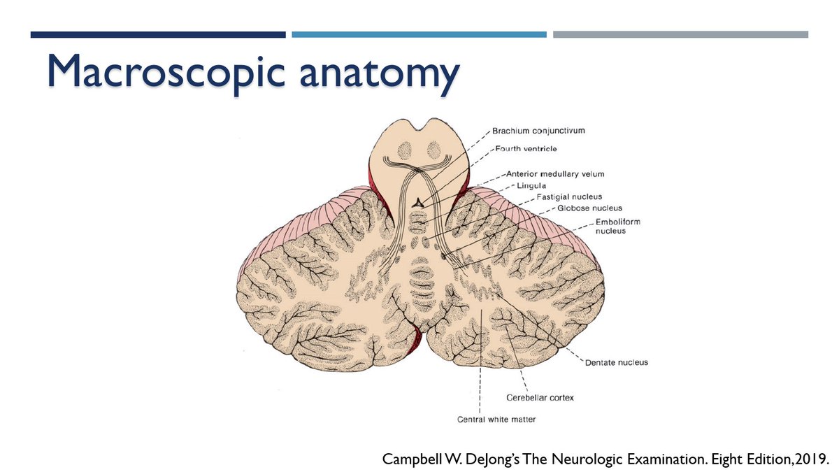
Deep nuclei: from lateral to medial.
-Dentate
-Emboliform + Globose = interposed
-Fastigial
"Don't Eat Greasy Foods" mnemonic some use to remember them.
13/🧵
-Dentate
-Emboliform + Globose = interposed
-Fastigial
"Don't Eat Greasy Foods" mnemonic some use to remember them.
13/🧵

Ready for the microscopic anatomy🔬⁉️
Here are some drawings by the great #RamónYCajal to relax before we continue.
14/🧵
Here are some drawings by the great #RamónYCajal to relax before we continue.
14/🧵

Microscopic anatomy:
Cortex ➡️ Three layers (from exterior to interior)
- Molecular
- Purkinje
- Granular
15/🧵
Cortex ➡️ Three layers (from exterior to interior)
- Molecular
- Purkinje
- Granular
15/🧵

Microscopic anatomy:
5⃣ Types of cells distributed in the three layers:
- Molecular: stellate and basket
- Purkinje cells: dendrites of these cells are in the molecular layer
- Granular: granule and Golgi cells
16/🧵
5⃣ Types of cells distributed in the three layers:
- Molecular: stellate and basket
- Purkinje cells: dendrites of these cells are in the molecular layer
- Granular: granule and Golgi cells
16/🧵

Microscopic anatomy:
2⃣ type of fibers arrive INTO the cerebellum and interact with the 5⃣ cells in the different layers.
Fibers come from tracts described in 10/
Final goal: reach molecular layer and synapse with Purkinje's cells dendrites.
17/🧵
2⃣ type of fibers arrive INTO the cerebellum and interact with the 5⃣ cells in the different layers.
Fibers come from tracts described in 10/
Final goal: reach molecular layer and synapse with Purkinje's cells dendrites.
17/🧵

Microscopic anatomy:
2⃣ Types of fibers
- Climbing fiber: signals from inf. Olivary Nucleus ➡️reach molecular layer and synapse with Purkinje's dendrites
18/🧵
2⃣ Types of fibers
- Climbing fiber: signals from inf. Olivary Nucleus ➡️reach molecular layer and synapse with Purkinje's dendrites
18/🧵

Microscopic anatomy:
2⃣ Types of fibers
- Mossy fibers: come from other tracts ➡️ Synapse in Granular layer (with granular cells) ➡️ Axons from the granular layer form the parallel fibers which synapse with dendrites of Purkinje's cells at molecular layer
19/🧵
2⃣ Types of fibers
- Mossy fibers: come from other tracts ➡️ Synapse in Granular layer (with granular cells) ➡️ Axons from the granular layer form the parallel fibers which synapse with dendrites of Purkinje's cells at molecular layer
19/🧵

Purkinje cells:
Are they importante? Yes
Really? Yes
Why? The carry signlas from the cerebellar CORTEX to the deep NUCLEI
And from there? To the rest of the CNS
Deep nuclei➡️ send signals outside the cerebellum to the rest of the CNS
20/🧵
Are they importante? Yes
Really? Yes
Why? The carry signlas from the cerebellar CORTEX to the deep NUCLEI
And from there? To the rest of the CNS
Deep nuclei➡️ send signals outside the cerebellum to the rest of the CNS
20/🧵

For details on clinical manifestations associated with cerebellar dysfunction I highly recomend this tweetorial (as well as part II) by the great @theneurolander
21/🧵
21/🧵
This next paper is amazing as well.
22/🧵
22/🧵

There is also Cognitive Dysfunction associated with Cerebellar lesions.
I think this is a great example of Brain Diaschisis or remote effects of a focal lesion on the function of
the brain.
23/🧵
I think this is a great example of Brain Diaschisis or remote effects of a focal lesion on the function of
the brain.
23/🧵


A few more final words:
Brain anatomy is complex, details come with time, repetition and practice. If we have the basis clear, the next steps will be much easier.
Hope you have enjoyed this brief introduction to cerebellar anatomy.
24/24
Brain anatomy is complex, details come with time, repetition and practice. If we have the basis clear, the next steps will be much easier.
Hope you have enjoyed this brief introduction to cerebellar anatomy.
24/24
Sources:
1.- Handb Clin Neurol. 2018;154:3-26.
doi: 10.1016/B978-0-444-63956-1.00001-1
2.- Neurol Clin. 2014 Nov;32(4):859-69.
doi: 10.1016/j.ncl.2014.07.01
3.- Handb Clin Neurol. 2018;154:85-108.
doi: 10.1016/B978-0-444-63956-1.00006-0
1.- Handb Clin Neurol. 2018;154:3-26.
doi: 10.1016/B978-0-444-63956-1.00001-1
2.- Neurol Clin. 2014 Nov;32(4):859-69.
doi: 10.1016/j.ncl.2014.07.01
3.- Handb Clin Neurol. 2018;154:85-108.
doi: 10.1016/B978-0-444-63956-1.00006-0
Sources:
4.- Blumenfeld H. Neuroanatomy Through Clinical Cases, 2010
5.- Front Syst Neurosci. 2021 Apr 9;15:646052.
doi: 10.3389/fnsys.2021.646052
6.- Board Review Series. Neuroanatomy. Fifth Ed. 2014
7.- Campbell W. DeJong’s The Neurologic Examination. Eight Edition,2019.
4.- Blumenfeld H. Neuroanatomy Through Clinical Cases, 2010
5.- Front Syst Neurosci. 2021 Apr 9;15:646052.
doi: 10.3389/fnsys.2021.646052
6.- Board Review Series. Neuroanatomy. Fifth Ed. 2014
7.- Campbell W. DeJong’s The Neurologic Examination. Eight Edition,2019.
Sources:
8.- Mayo Clinic Neurology Board Review. 2015, Oxford Scientific press
9.- Cerebellum. 2016 Jun;15(3):369-91.
doi: 10.1007/s12311-015-0687-3
10.- Cerebellum. 2019 Oct;18(5):941-950.
doi: 10.1007/s12311-019-01060-2
8.- Mayo Clinic Neurology Board Review. 2015, Oxford Scientific press
9.- Cerebellum. 2016 Jun;15(3):369-91.
doi: 10.1007/s12311-015-0687-3
10.- Cerebellum. 2019 Oct;18(5):941-950.
doi: 10.1007/s12311-019-01060-2
Sources:
11.- Brain. 2018 Jan 1;141(1):248-270.
doi: 10.1093/brain/awx317
🤪
11.- Brain. 2018 Jan 1;141(1):248-270.
doi: 10.1093/brain/awx317
🤪
جاري تحميل الاقتراحات...