

Definition:
COPD is a chronic pulmonary disease characterized by persistent respiratory symptoms and airflow limitation.
Note 📝 COPD is irreversible.
COPD is a chronic pulmonary disease characterized by persistent respiratory symptoms and airflow limitation.
Note 📝 COPD is irreversible.
Etiology:
-The main cause is smoking
-environmental exposure (pollution and dust...)
-genetic (alpha-1 anti-trypsin deficiency)
-The main cause is smoking
-environmental exposure (pollution and dust...)
-genetic (alpha-1 anti-trypsin deficiency)

Theres two main conditions:
1-Chronic bronchitis
2-Emphysema
1-Chronic bronchitis
2-Emphysema
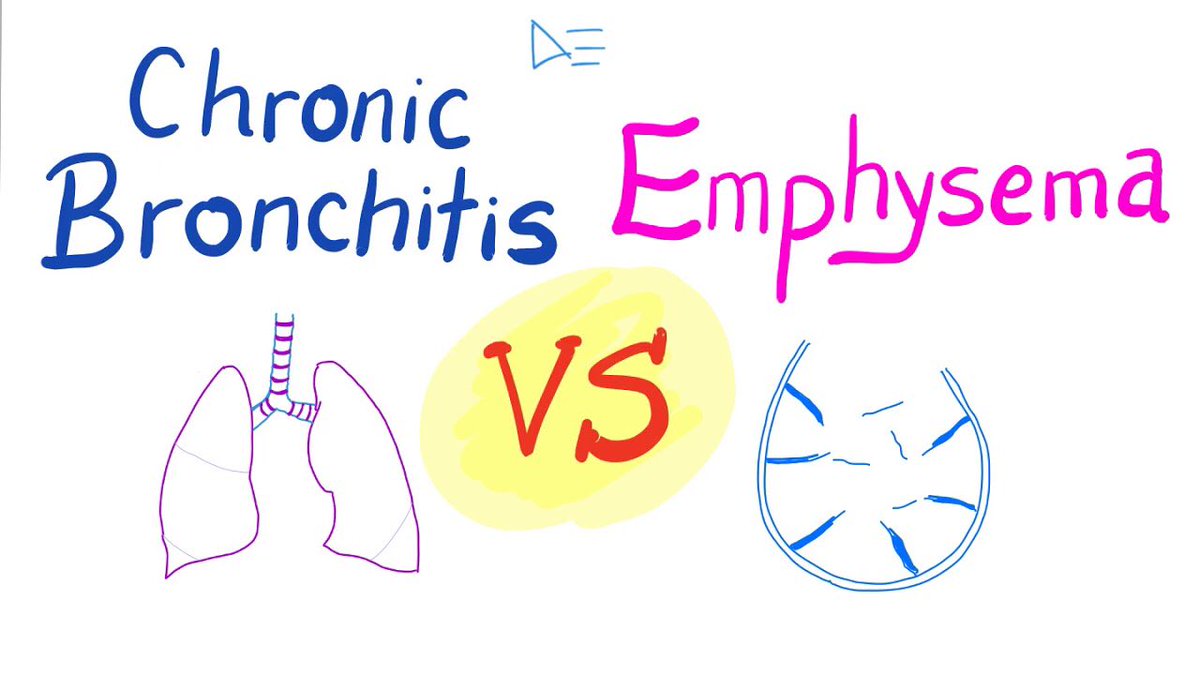
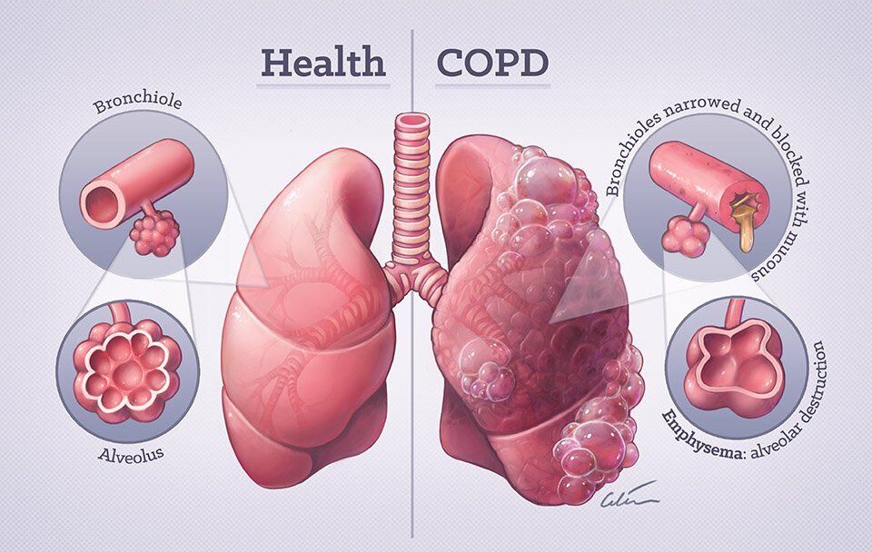
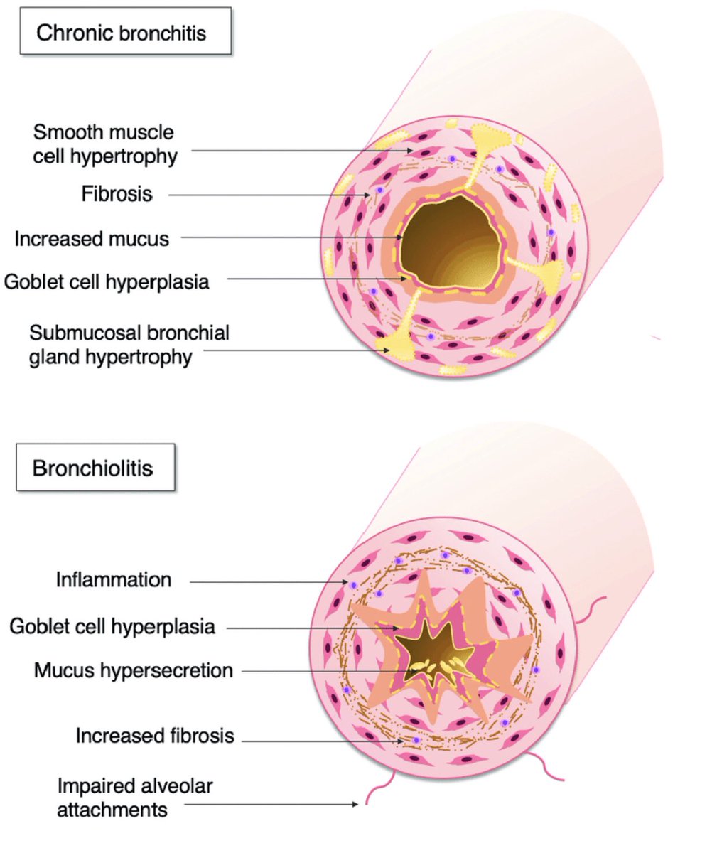
Chronic bronchitis:
Is mucous build up
Etiology:
Normally sub-mucosa glands and goblet cells release mucous to protect airway.
In this condition it may increase by smoking (hypertrophy and hyperplasia) and causing narrowing of airway lead to “ciliary dysfunction”➡️ air trapping.
Is mucous build up
Etiology:
Normally sub-mucosa glands and goblet cells release mucous to protect airway.
In this condition it may increase by smoking (hypertrophy and hyperplasia) and causing narrowing of airway lead to “ciliary dysfunction”➡️ air trapping.
Lead to increase CO2 in blood (hypercapnia)
And decrease O2 in blood(hypoxemia).
Note 📝 mucous building up is risk factor of pneumonia.
And decrease O2 in blood(hypoxemia).
Note 📝 mucous building up is risk factor of pneumonia.
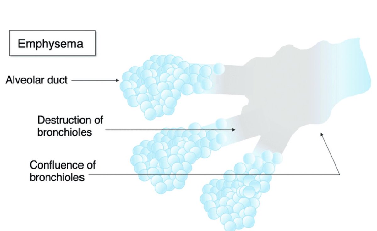
Emphysema:
disorder affecting the alveoli (tiny air sacs) of the lungs. The transfer of oxygen and carbon dioxide in the lungs takes place in the walls of the alveoli. In emphysema, the alveoli become abnormally inflated, damaging their walls and making it harder to breathe.
disorder affecting the alveoli (tiny air sacs) of the lungs. The transfer of oxygen and carbon dioxide in the lungs takes place in the walls of the alveoli. In emphysema, the alveoli become abnormally inflated, damaging their walls and making it harder to breathe.
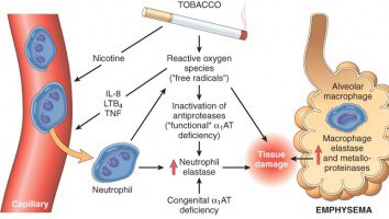
Etiology:
Smoke or dust make cytokines to activate neutrophils to release protases specially elastase that break elastin.
Elastin function is to keep air way open.
Smoke or dust make cytokines to activate neutrophils to release protases specially elastase that break elastin.
Elastin function is to keep air way open.
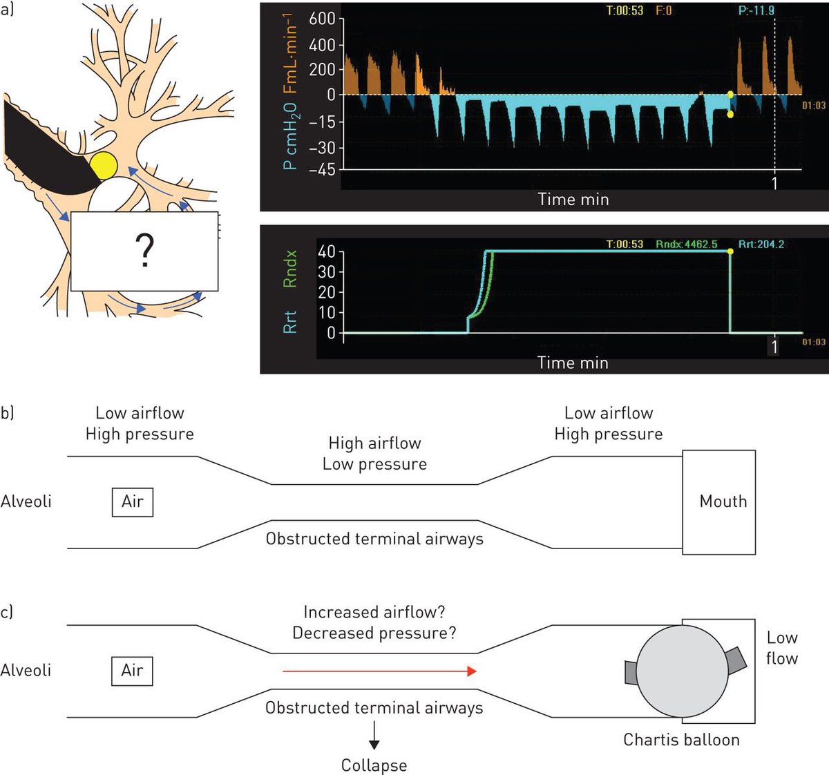
Bernoulli principle:
Low pressure inside alveoli
High pressure outside
Elastic prevent alveoli from collapsing
If elastic damaged ➡️alveoli collapse and lead to air trapping.
Low pressure inside alveoli
High pressure outside
Elastic prevent alveoli from collapsing
If elastic damaged ➡️alveoli collapse and lead to air trapping.
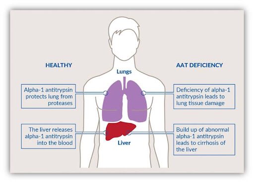
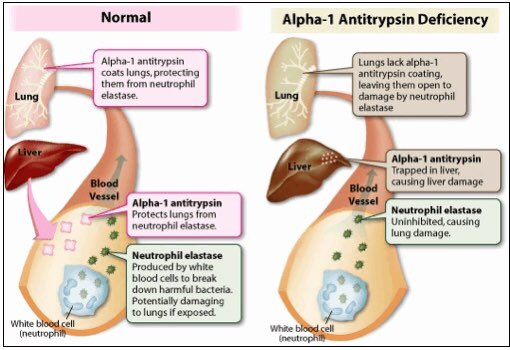
Note 📝 liver release alpha anti trypsin this inhibit the elastase.
So in pt who has alpha anti trypsin deficiency this process can’t happened.
So in pt who has alpha anti trypsin deficiency this process can’t happened.
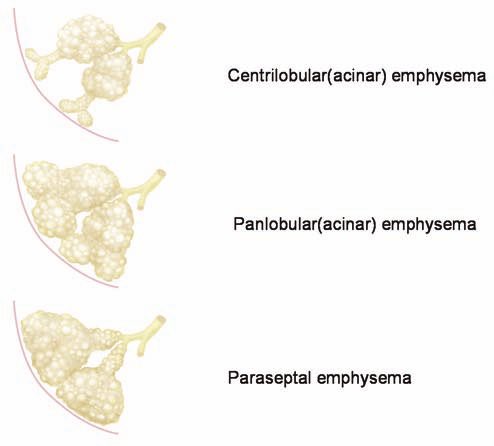
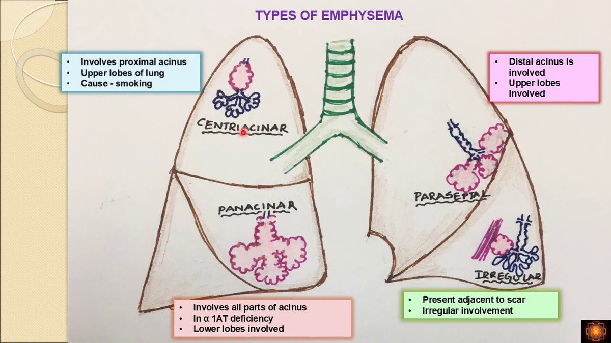
Types of emphysema:
-centriacinar➡️ in proximal airways (upper lopes).
-panacinar ➡️ in distal airways (lower lopes).
-paraseptal or disalacinar ➡️ near to pleura.
Note 📝 paraseptal may lead to close pneumothorax.
-centriacinar➡️ in proximal airways (upper lopes).
-panacinar ➡️ in distal airways (lower lopes).
-paraseptal or disalacinar ➡️ near to pleura.
Note 📝 paraseptal may lead to close pneumothorax.
cor pulmonale:
When the ventilation is low the perfusion become low, HOW?
By hypoximic vasoconstriction ➡️ pulmonary HTN ➡️ right ventricle hypertrophy ➡️ right side heart failure.
When the ventilation is low the perfusion become low, HOW?
By hypoximic vasoconstriction ➡️ pulmonary HTN ➡️ right ventricle hypertrophy ➡️ right side heart failure.

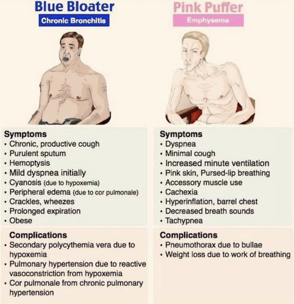
Signs and symptoms:
blue bloater (chronic bronchitis)
vs
pink puffer (emphysema)
blue bloater (chronic bronchitis)
vs
pink puffer (emphysema)
Diagnosis:
1-clinical diagnosis
Chronic bronchitis (3months/yr x 2yrs) of productive cough.
2-pulmonary function tests:
PFTs <75% ➡️ obstructive issue.
FVC low
FEV1 very low
So bronchodilator given like SABA then FEV1 increase 12% ➡️ sign of COPD.
1-clinical diagnosis
Chronic bronchitis (3months/yr x 2yrs) of productive cough.
2-pulmonary function tests:
PFTs <75% ➡️ obstructive issue.
FVC low
FEV1 very low
So bronchodilator given like SABA then FEV1 increase 12% ➡️ sign of COPD.
3-pulse oximeter:
<88%
Or
<90% with HF or polycythemia
4- ABG:
⬆️PCO2 ⬇️PH ⬇️Po2
5-ECG:
-Multifocal atrial tachycardia seen in COPD pt
-right ventricle hypertrophy
6-CXR:
Air trapping
Flat diaphragm
Increase anterposterior AP diameter.
<88%
Or
<90% with HF or polycythemia
4- ABG:
⬆️PCO2 ⬇️PH ⬇️Po2
5-ECG:
-Multifocal atrial tachycardia seen in COPD pt
-right ventricle hypertrophy
6-CXR:
Air trapping
Flat diaphragm
Increase anterposterior AP diameter.
Note 📝 emphysema (decrease vascular markings).
Chronic bronchitis (increase vascular markings).
Chronic bronchitis (increase vascular markings).
Treatment:
-smoking cessation
-influenza and pneumococcal vaccine
-supplemental oxygen between 88-92%
-Bronchodilators:
Mild or intermittent (SAMA + SABA medium dose inhaler or nebulizer).
Moderate or sever FEV1 <60% and still symptomatic
So we use(LAMA + LABA).
-smoking cessation
-influenza and pneumococcal vaccine
-supplemental oxygen between 88-92%
-Bronchodilators:
Mild or intermittent (SAMA + SABA medium dose inhaler or nebulizer).
Moderate or sever FEV1 <60% and still symptomatic
So we use(LAMA + LABA).
Acute exacerbation of COPD:
Corticosteroids IV or PO
Long term antibiotic (azithromycin) may given.
Corticosteroids IV or PO
Long term antibiotic (azithromycin) may given.
جاري تحميل الاقتراحات...