

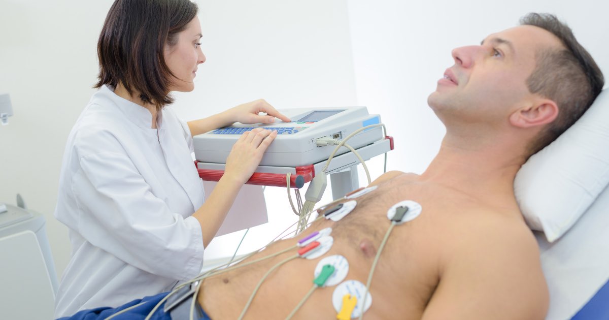
Electrocardiography is the process of producing an electrocardiogram. It is a graph of voltage versus time of the electrical activity of the heart using electrodes placed on the skin.
Note 📝 Cardiac tissues consist of:
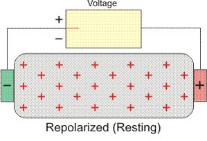
-positive electrodes
-negative electrodes
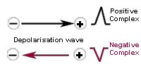
When the electrical activity moving from negative to positive electrode ➡️ positive deflection.
When the electrical activity moving from positive to negative electrode ➡️ positive deflection.
-positive electrodes
-negative electrodes
When the electrical activity moving from negative to positive electrode ➡️ positive deflection.
When the electrical activity moving from positive to negative electrode ➡️ positive deflection.
Note 📝 when the electrical activity moving too slow ➡️flat line/ isoelectric line
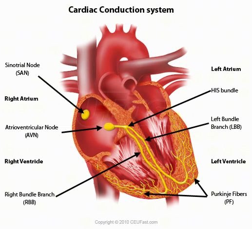
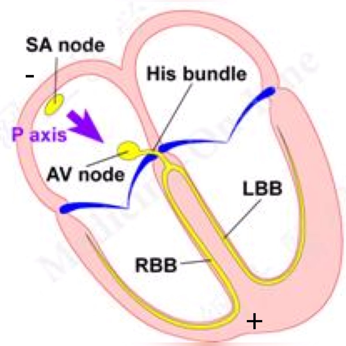
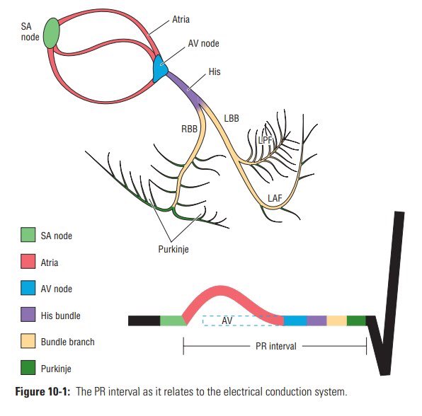
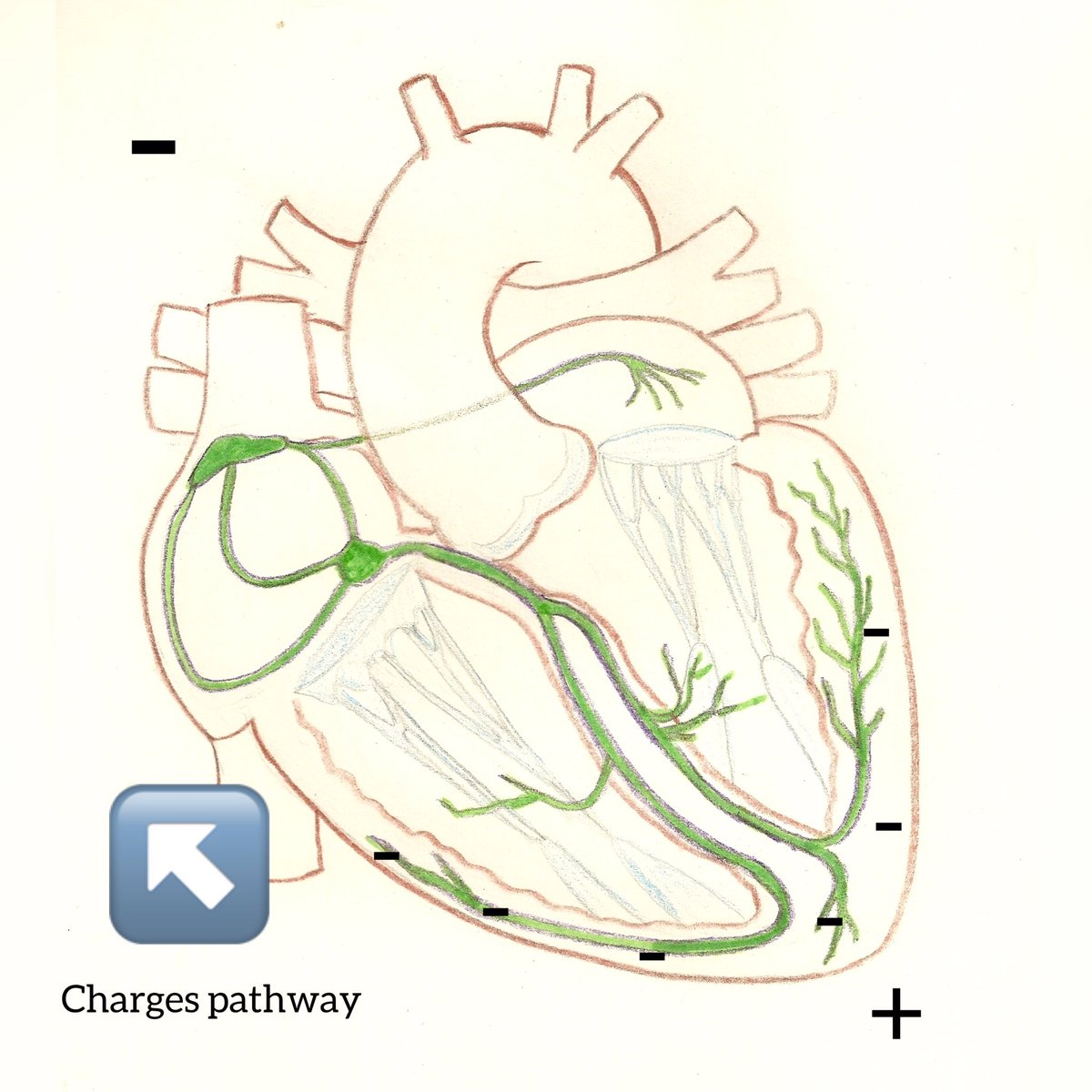
Cardiac conduction system:
SA node pacemaker
AV node
Bundle of HIS
left and right bundle branch
Purkinje fibers
SA node pacemaker
AV node
Bundle of HIS
left and right bundle branch
Purkinje fibers
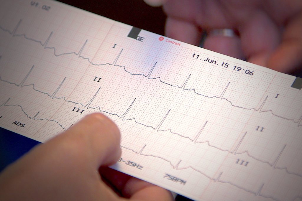
How to read ECG on lead II:
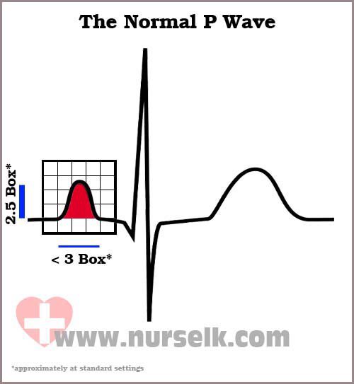
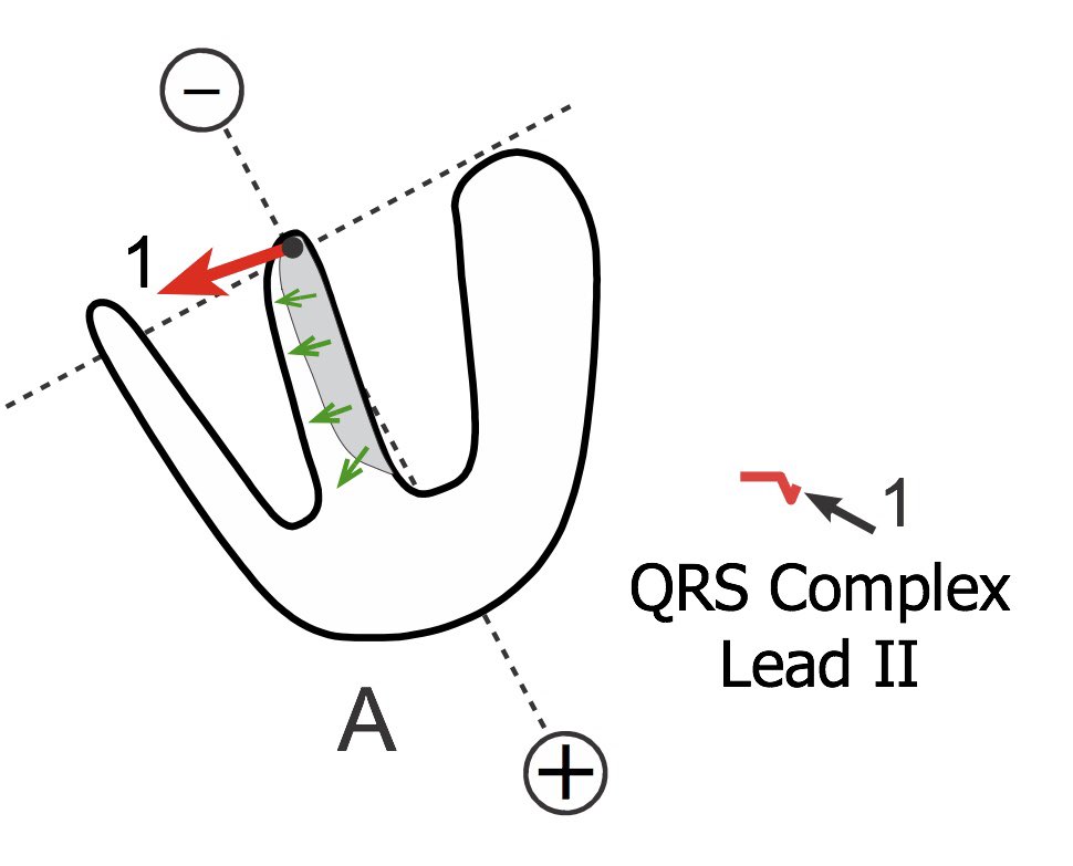
1- P wave
The P wave represents the wave of depolarization that spreads from the SA node throughout the atria, and is usually 0.08 to 0.10 seconds (80-100 ms) in duration.
➡️Positive deflection
Called atrial depolarization
1- P wave
The P wave represents the wave of depolarization that spreads from the SA node throughout the atria, and is usually 0.08 to 0.10 seconds (80-100 ms) in duration.
➡️Positive deflection
Called atrial depolarization
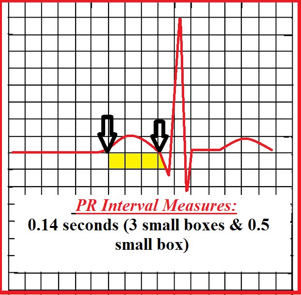
2- P-R interval
is the time from the beginning of the P wave (atrial depolarization) to the beginning of the QRS complex (ventricular depolarization). The normal PR interval measures 0.12-0.20 seconds
AV node slow conductor (0.1 sec).
It’s important to identify AV blocks.
is the time from the beginning of the P wave (atrial depolarization) to the beginning of the QRS complex (ventricular depolarization). The normal PR interval measures 0.12-0.20 seconds
AV node slow conductor (0.1 sec).
It’s important to identify AV blocks.
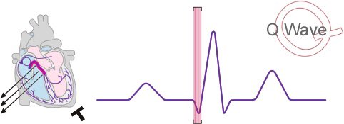
3- Q wave
Action potential conducted through AV node to the right and left bundle branch.
Left branch is the main cause of septum depolarization so this cause negative deflection.
Form positive to negative (negative deflection).
The duration of the Q waves is 0.03 second or less
Action potential conducted through AV node to the right and left bundle branch.
Left branch is the main cause of septum depolarization so this cause negative deflection.
Form positive to negative (negative deflection).
The duration of the Q waves is 0.03 second or less
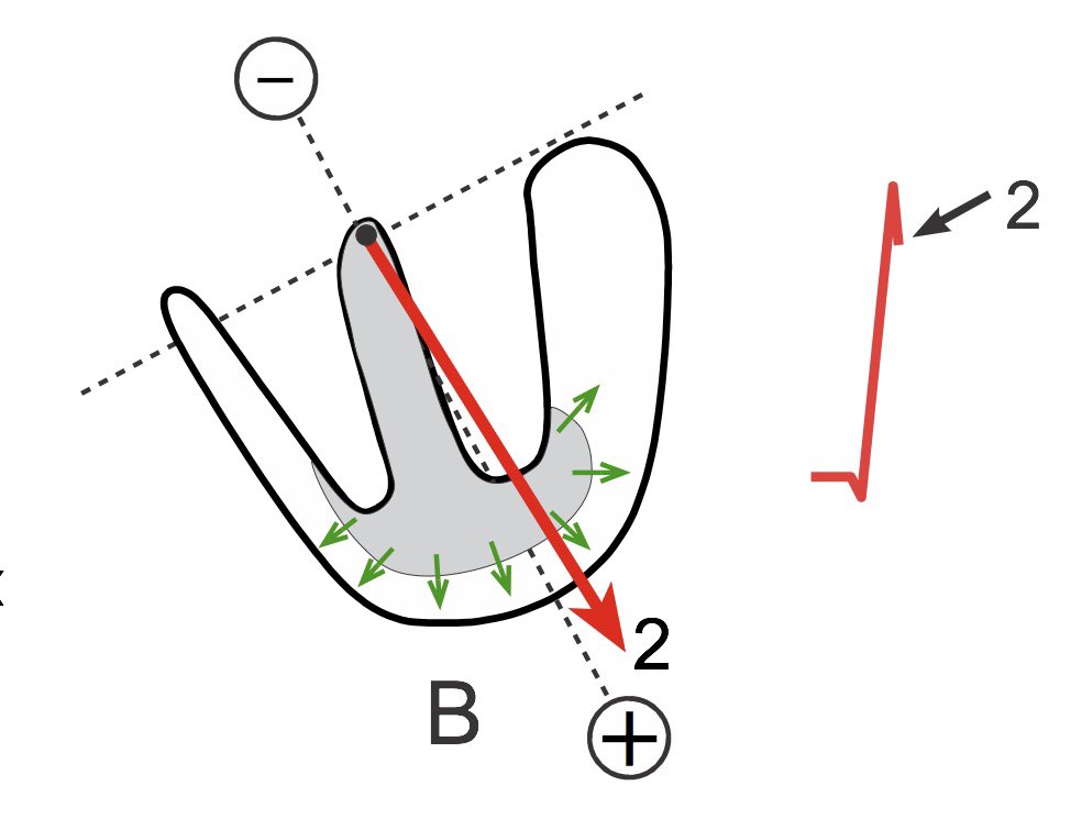
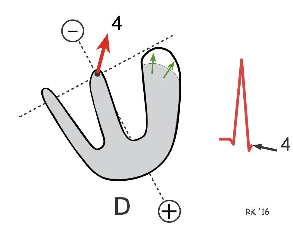
4- R wave
Action potentials goes to bundle of His.
positive deflection.
Note 📝 left ventricle thicker than right ventricle, so it’s generate more electricity
Action potentials goes to bundle of His.
positive deflection.
Note 📝 left ventricle thicker than right ventricle, so it’s generate more electricity
5- S wave
When electrical activities moving toward to the bases of ventricles (depolarization bases of ventricles).
When electrical activities moving toward to the bases of ventricles (depolarization bases of ventricles).
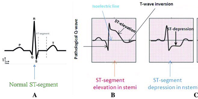
6- S-T segment
ST Segment represents the interval between ventricular depolarization and repolarization.
Isoelectric line.
The most important cause of ST segment abnormality (elevation or depression) is myocardial ischaemia or infarction.
duration is usually around 0.08 sec.
ST Segment represents the interval between ventricular depolarization and repolarization.
Isoelectric line.
The most important cause of ST segment abnormality (elevation or depression) is myocardial ischaemia or infarction.
duration is usually around 0.08 sec.
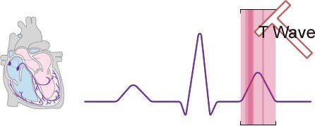
7- T wave
When the charges turns negative from positive charges and it’s moves away from positive, so lead to positive deflection
When the charges turns negative from positive charges and it’s moves away from positive, so lead to positive deflection
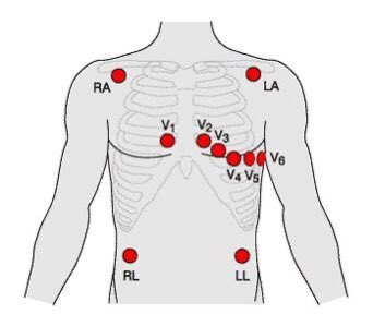
12 leads ECG:
⁃3 bipolar (I, II, III).
⁃3 augmented (aVF, aVR, aVL).
⁃6 chest leads (V1-V6).
Note 📝 bipolar and augmented leads pics anterior and posterior of the heart.
Chest lead pics superior and inferior of the heart.
⁃3 bipolar (I, II, III).
⁃3 augmented (aVF, aVR, aVL).
⁃6 chest leads (V1-V6).
Note 📝 bipolar and augmented leads pics anterior and posterior of the heart.
Chest lead pics superior and inferior of the heart.
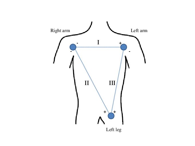
Bipolar leads:
Lead I ➡️ looking to the heart from left lateral side
Lead II and lead III ➡️ looking to the heart from the bottom
Note 📝 in the pic, + charge like your eye when you start looking.
Lead I ➡️ looking to the heart from left lateral side
Lead II and lead III ➡️ looking to the heart from the bottom
Note 📝 in the pic, + charge like your eye when you start looking.
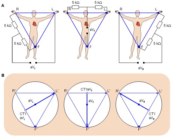
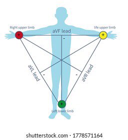
Augmented leads:
aVR➡️ looking to the heart form right side
aVF ➡️ looking to the heart from bottom
aVL ➡️ looking to the heart from left side
aVR➡️ looking to the heart form right side
aVF ➡️ looking to the heart from bottom
aVL ➡️ looking to the heart from left side
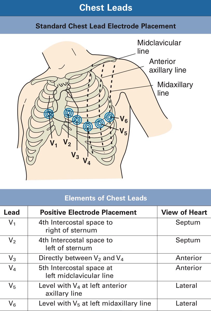
Chest leads:
V1 and V2 ➡️ take pic of septum or posterior
V3 and V4 ➡️ take pic of anterior wall of heart
V5 and V6 ➡️ take pic of left lateral wall of heart
V1 and V2 ➡️ take pic of septum or posterior
V3 and V4 ➡️ take pic of anterior wall of heart
V5 and V6 ➡️ take pic of left lateral wall of heart
وبكذا انتهينا من الجزء الاول وبإذن الله الثريد القادم راح يكون تكمله لهذا الثريد 🙏❤️
تعديل لخطأ
When the electrical activity moving from positive to negative electrode ➡️negative deflection.
When the electrical activity moving from positive to negative electrode ➡️negative deflection.
جاري تحميل الاقتراحات...