

Don't let your patients die from AAA or Aortic Dissection! #POCUS
1⃣Learn how to Easily do Aorta Ultrasound
2⃣Learn how to perform the Suprastenral View
3⃣Recognize AAA and Aortic Dissection
✅New Blog Post 👉🔗pocus101.com
#medtweetorial👇(1/25)
1⃣Learn how to Easily do Aorta Ultrasound
2⃣Learn how to perform the Suprastenral View
3⃣Recognize AAA and Aortic Dissection
✅New Blog Post 👉🔗pocus101.com
#medtweetorial👇(1/25)
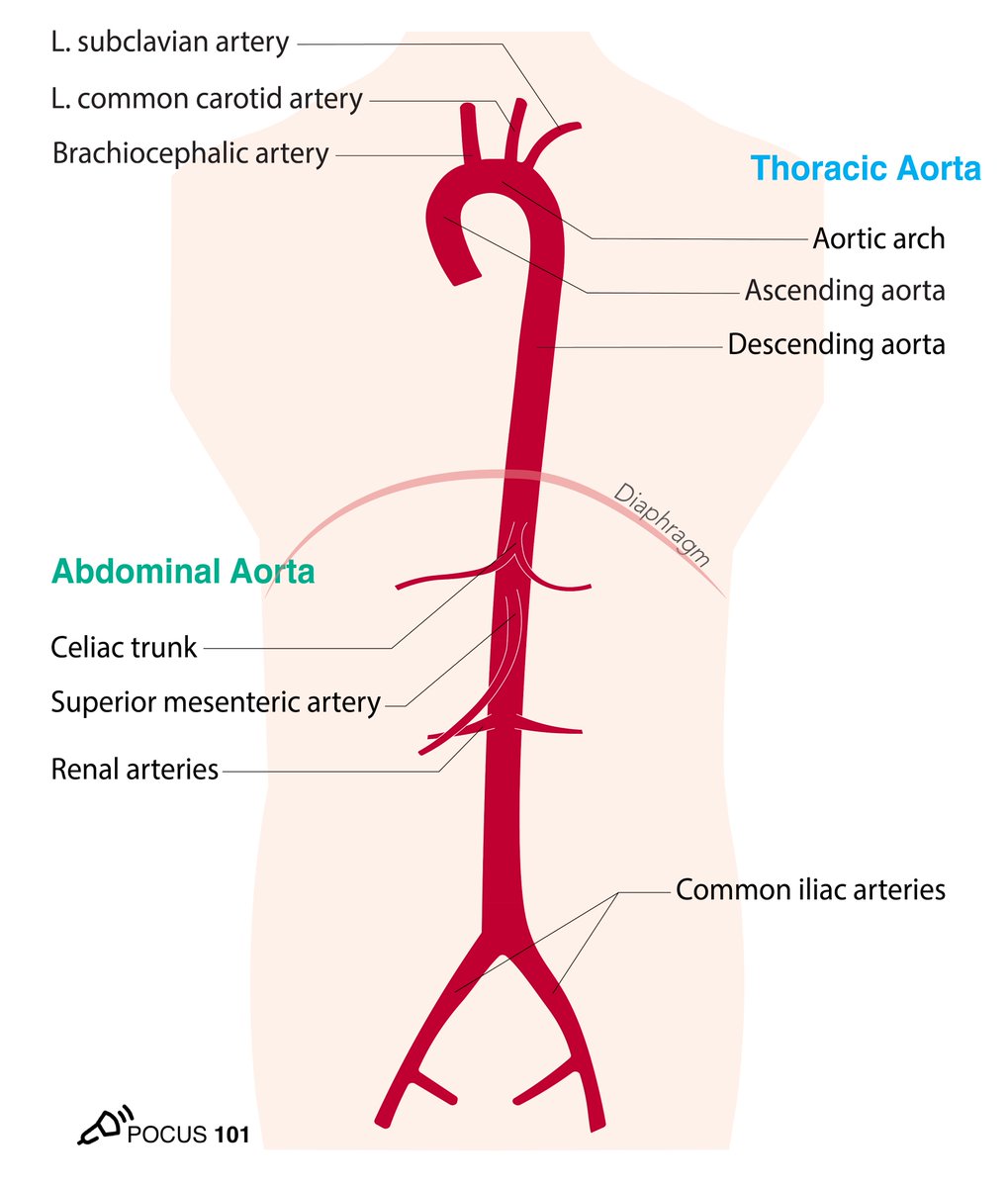

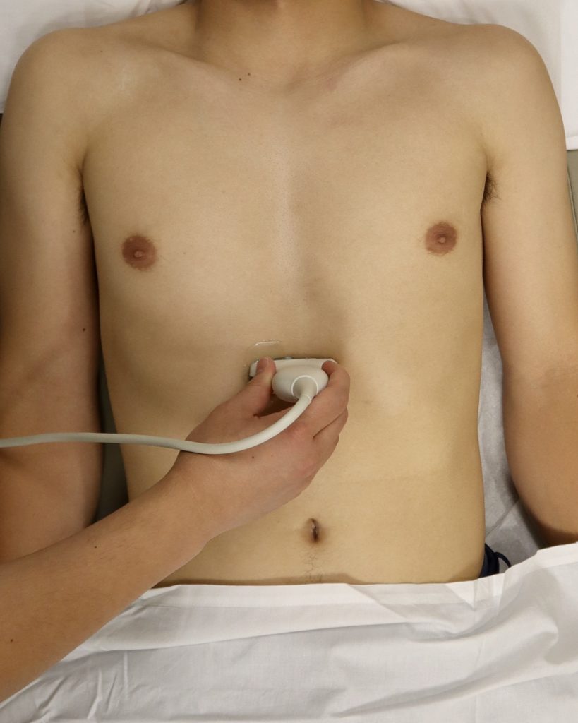
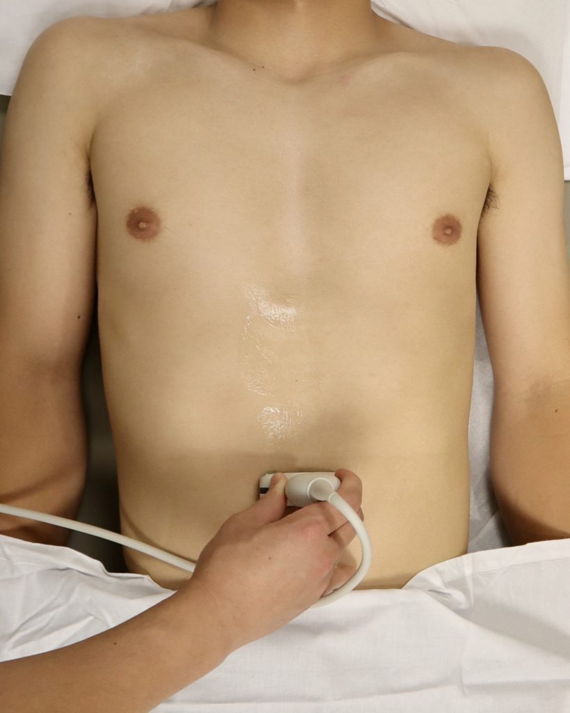
2 Start with the Short axis approach and place the probe near the subxiphoid area
👉🔗pocus101.com
👉🔗pocus101.com


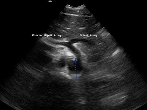
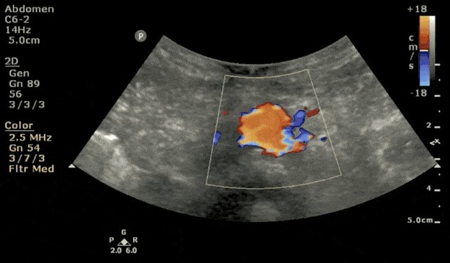
4 Slide your probe a little more inferiorly to see the "Seagull Sign": Celiac Trunk, Splenic Artery, and Common Hepatic Artery. Using Color Doppler may help!
👉🔗pocus101.com
👉🔗pocus101.com
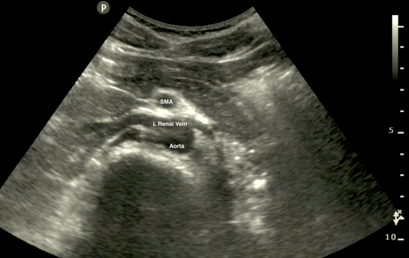
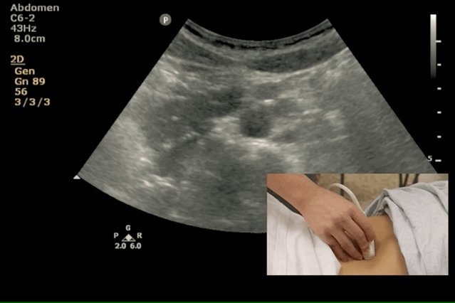
5 As you slide more inferiorly you should be able to see the Superior Mesenteric Artery (SMA). Occasionally you will see the Left Renal Vein between the SMA and the Aorta.
👉🔗pocus101.com
👉🔗pocus101.com
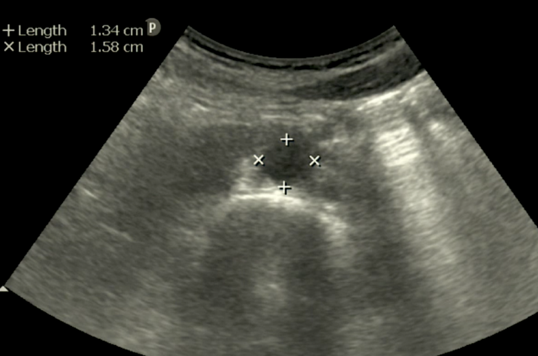
6 Measure the abdominal aorta at it's widest diameter from Outer to Outer walls. Obtain both an anteroposterior measurement and a transverse measurement if possible.
👉🔗pocus101.com
👉🔗pocus101.com
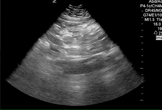
7 Next move on to the Mid Aorta by continuing to slide your probe inferiorly.
👉🔗pocus101.com
👉🔗pocus101.com


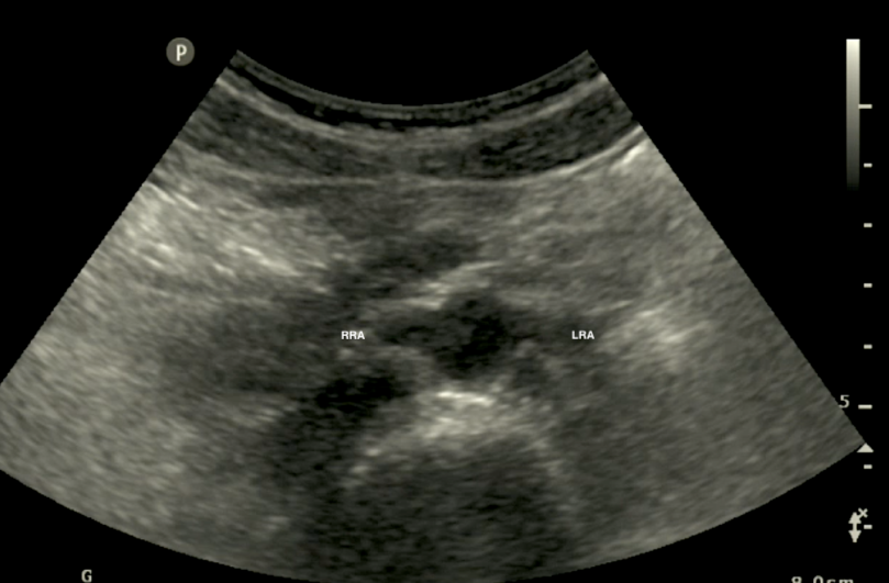
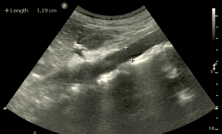
8 Take a measurement of the aorta in the Mid Aorta. Sometimes you may even see the right and left renal arteries.
👉🔗pocus101.com
👉🔗pocus101.com
9 Continue to slide your probe inferiorly to visualize the distal aorta. You may need to decrease your depth as it is usually the most superficial part of the aorta.
👉🔗pocus101.com
👉🔗pocus101.com
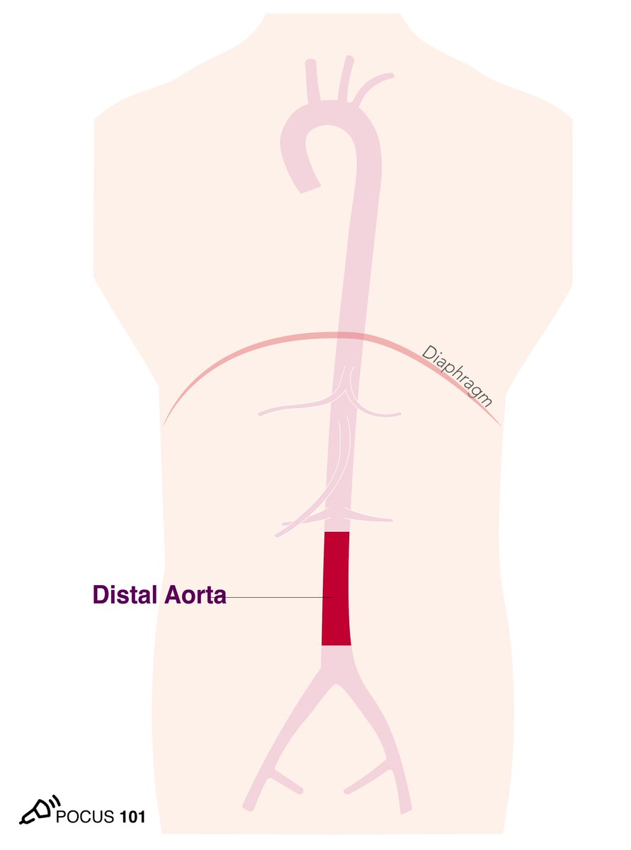
10 Scan all the way to the iliac bifurcation and take a measurement of the distal aorta before the bifurcation.
👉🔗pocus101.com
👉🔗pocus101.com
11 Obtain a long axis view of the aorta by bringing the probe back to the epigastric area and from the short axis view, rotate the probe 90 degrees clockwise.
👉🔗pocus101.com
👉🔗pocus101.com
12 Visualize the Celiac Trunk and the SMA coming off the aorta in long axis.
👉🔗pocus101.com
👉🔗pocus101.com

13 Measure the anteroposterior diameter. Of note the short axis view is more reliable than the long axis view for accurate measurements.
👉🔗pocus101.com
👉🔗pocus101.com
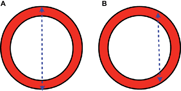
14 Cylinder Tangent Effect: Panel (A) shows an ultrasound measurement through the center of the aorta will provide the maximal AP diameter. Panel (B) shows a measurement to the side (tangent) of the aorta resulting in a falsely small aortic diameter.
👉🔗pocus101.com
👉🔗pocus101.com
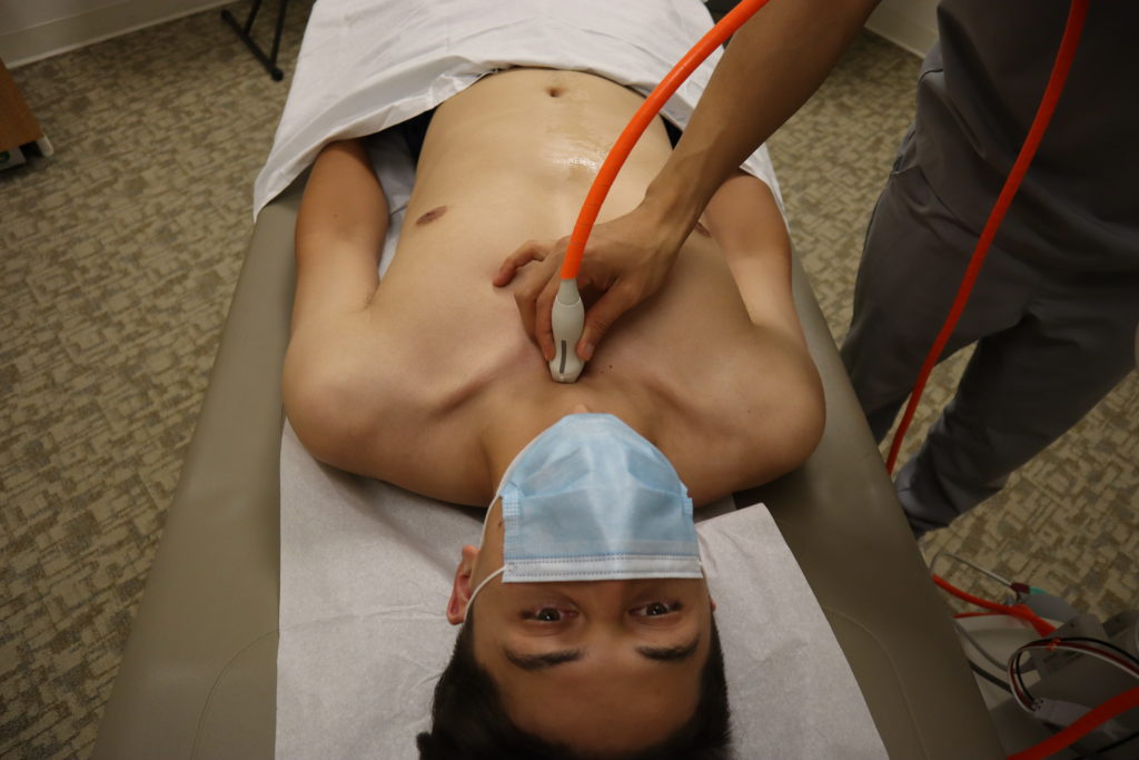
15 Obtain a suprasternal notch view
1) Place the indicator towards the patient’s head.
2) Rock the tail of the probe superiorly.
3) Rotate slightly clockwise (15-20˚) towards the patient’s left shoulder.
👉🔗pocus101.com
1) Place the indicator towards the patient’s head.
2) Rock the tail of the probe superiorly.
3) Rotate slightly clockwise (15-20˚) towards the patient’s left shoulder.
👉🔗pocus101.com
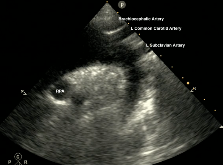
16 Identify the Aortic Arch, Brachycephalic Artery, Left Common Carotid Artery, Left Subclavian Artery and Right Pulmonary Artery (RPA).
👉🔗pocus101.com
👉🔗pocus101.com
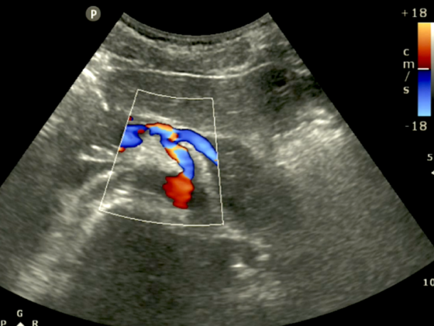
17 Apply color Doppler to the Suprasternal notch view. The ascending aorta should have red flow (towards probe) and the descending aorta should have blue flow (away from probe)
👉🔗pocus101.com
👉🔗pocus101.com
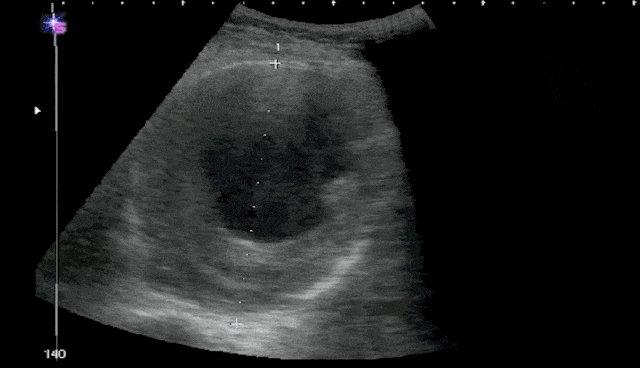
18 Fusiform AAA's are symmetrical, circumferential dilatations of the aorta.
👉🔗pocus101.com
👉🔗pocus101.com
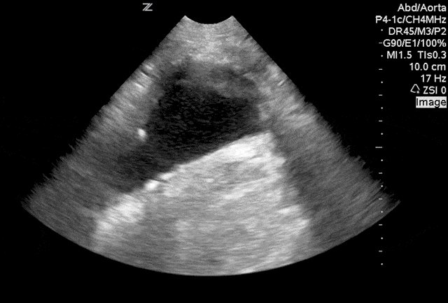
19 Saccular aneurysms form as asymmetrical outpocketings of the aortic wall.
👉🔗pocus101.com
👉🔗pocus101.com
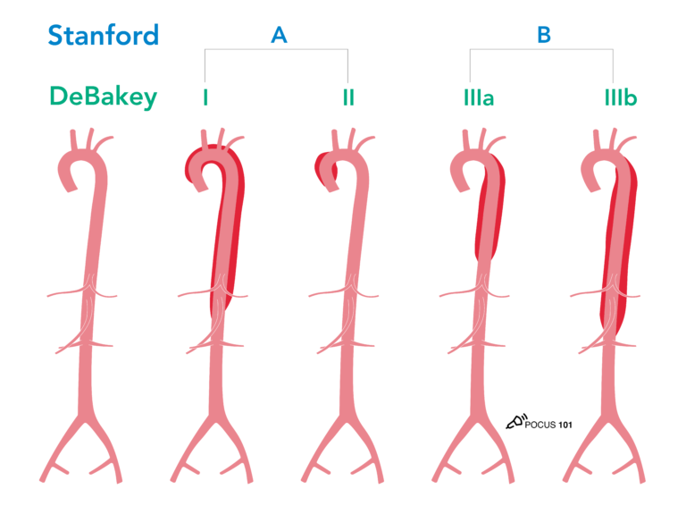
20 Recall the Stanford vs Debakey Classifications for Aortic Dissection. Use ALL the views you can including TTE, aorta ultrasound, and suprasternal views.
Remember, however, TEE and CT angio are the gold standard.
👉🔗pocus101.com
Remember, however, TEE and CT angio are the gold standard.
👉🔗pocus101.com
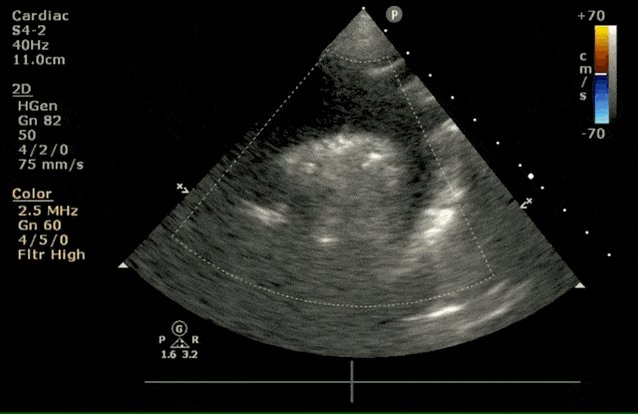
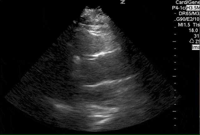
21 This transthoracic parasternal long axis view shows a echogenic dissection flap just distal to the aortic valve.
👉🔗pocus101.com
👉🔗pocus101.com
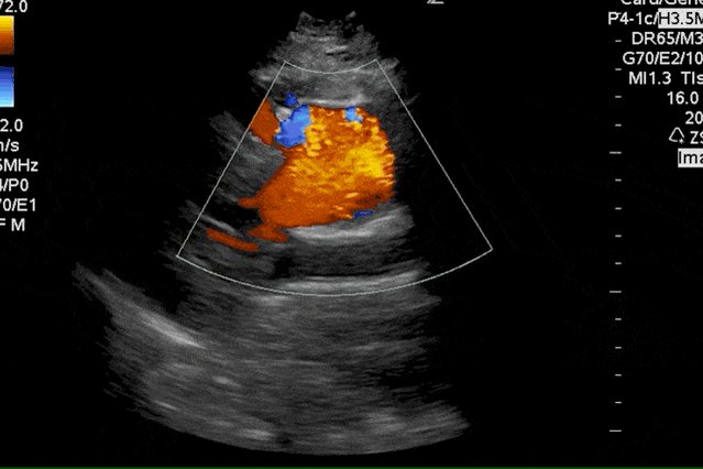
22 This suprasternal notch view shows an aortic dissection involving the aortic arch and the brachiocephalic and left common carotid artery. This particular patient presented with unilateral right sided weakness.
👉🔗pocus101.com
👉🔗pocus101.com
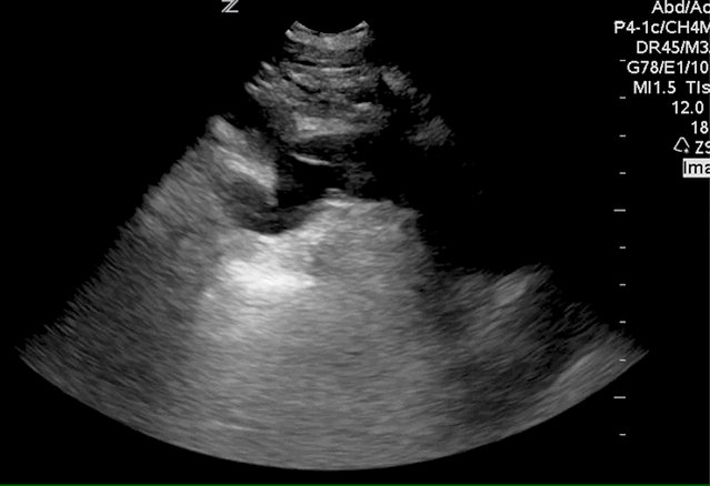
23 Aorta ultrasound, is a quick way to see if the aortic dissection involves the abdominal aorta.
👉🔗pocus101.com
👉🔗pocus101.com
24 Aortic regurgitation in aortic dissection will be seen as retrograde diastolic blood flow from the aortic arch, seen with Doppler. This is what causes your diastolic murmur!
👉🔗pocus101.com
👉🔗pocus101.com
25 Want to learn more about Aorta Ultrasound? Check out our new #POCUS Review book with hundreds of clinical ultrasound cases and a 20% discount code!
👉🔗pocus101.com
👉🔗pocus101.com

جاري تحميل الاقتراحات...